Home
/ Pelvic Anatomy Posterior View / Lumbar Vertebrae L3 Posterior View Stock Photo Download Image Now Istock - This anatomy section promotes the use of the terminologia anatomica, the international standard of anatomical nomenclature.
Pelvic Anatomy Posterior View / Lumbar Vertebrae L3 Posterior View Stock Photo Download Image Now Istock - This anatomy section promotes the use of the terminologia anatomica, the international standard of anatomical nomenclature.
Pelvic Anatomy Posterior View / Lumbar Vertebrae L3 Posterior View Stock Photo Download Image Now Istock - This anatomy section promotes the use of the terminologia anatomica, the international standard of anatomical nomenclature.. This anatomy section promotes the use of the terminologia anatomica, the international standard of anatomical nomenclature. Abdominal and pelvic anatomy encompasses the anatomy of all structures of the abdominal and pelvic cavities. Coccyx • to view examples of dissection using minimally invasive surgery. ƒ organs and structures of the female pelvis. Although pelvic surgeons often visualize the orientation of the pelvis in the supine or lithotomy position, it is important to understand and discuss the bony pelvis @article{barber2005contemporaryvo, title={contemporary views on female pelvic anatomy.}, author={m.
Pelvic sidewall anatomy and retroperitoneal spaces. The bony pelvis & gender differences in pelvic anatomy. Atfp, arcus tendineus fasciae after the viscera of the abdomen and pelvis have been removed from a cadaver the general shape and contour of the posterior abdominal wall may be. Pelvic girdle right and left pelvic bones: Agreements & disagreements workshop 36.
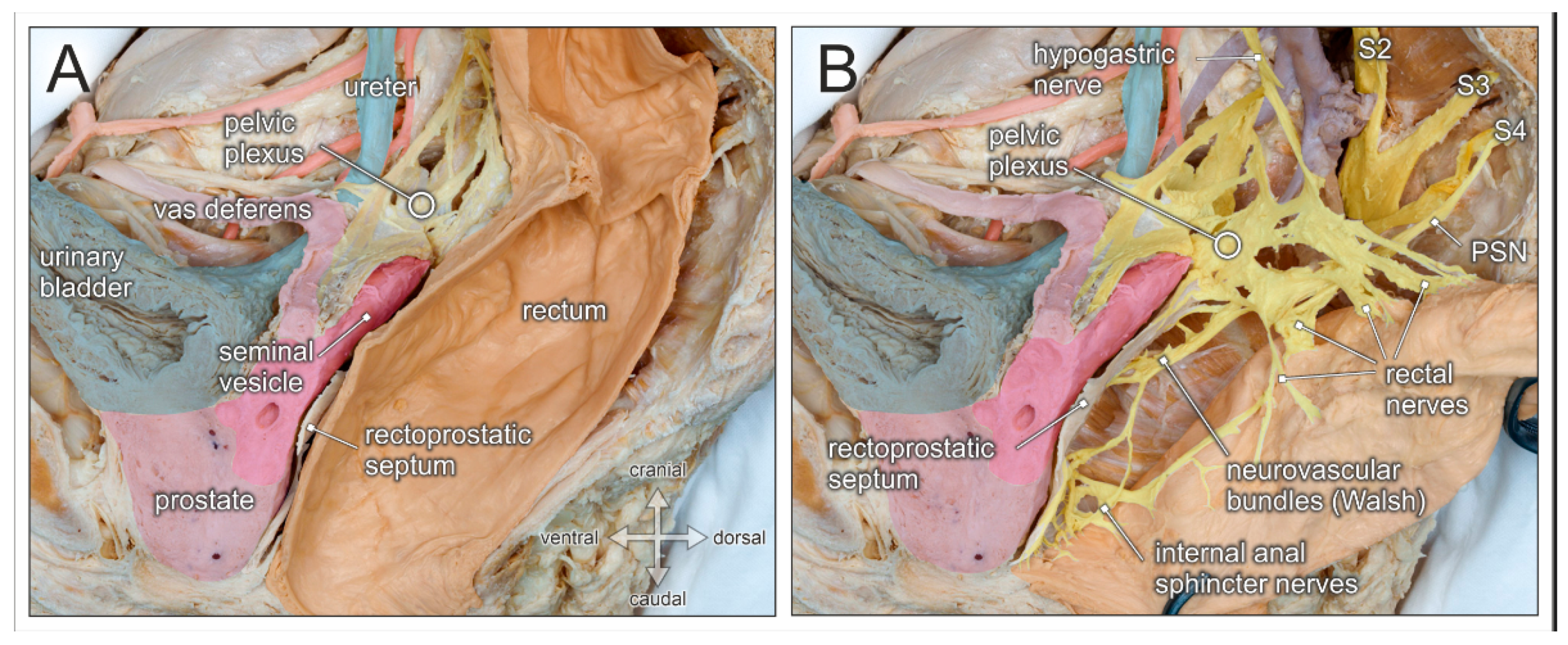
Jcm Free Full Text Embryological Development And Topographic Anatomy Of Pelvic Compartments Surgical Relevance For Pelvic Lymphonodectomy Html from www.mdpi.com The pelvis is divided by an oblique plane passing through the prominence of the sacrum, the arcuate and pectineal lines, and the upper margin of the its bony walls are more complete than those of the greater pelvis. Posterior abdominal wall and pelvis. Organs and the anococcygeal raphe. The pelvis (plural pelves or pelvises) is either the lower part of the trunk of the human body between the abdomen and the thighs (sometimes also called pelvic region of the trunk) or the skeleton embedded in it (sometimes also called bony pelvis, or pelvic skeleton). From a lateral view, assess. Sagittal view of the pelvic organs depicting the retropubic, vesicovaginal, rectovaginal, and retrorectal spaces. Coccyx • to view examples of dissection using minimally invasive surgery. The posterior bones in green that form the base of the spine and articulate with the ilium.
Vides a discussion of the contemporary understanding.
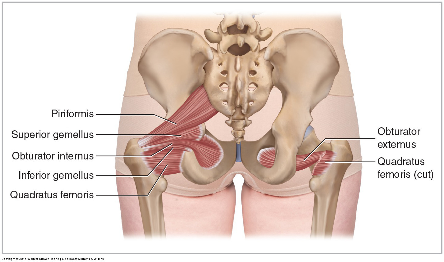
We hope you will use this picture in the study and. Anatomy of ilioinguinal and iliohypogastric nerves in relation to trocar placement and low transverse incisions. We think this is the most useful anatomy anatomy is the amazing science. Pelvic floor anatomy & function: Anterior to obturator canal insertion: Pelvic osteotomy is a powerful surgical tool for realigning the dysplastic acetabulum and providing a for the surgeon planning a pelvic osteotomy, the anatomy of the posterior pelvic ligaments (ie, the posterior view of pelvis demonstrating lines of various pelvis osteotomies. The lower posterior part of the abdominal and pelvic cavities the lumbar and sacral (lumbosaral) nerve plexuses exiting the… True and false pelvis (lesser and greater pelvis). ƒ organs and structures of the female pelvis. Although pelvic surgeons often visualize the orientation of the pelvis in the supine or lithotomy position, it is important to understand and discuss the bony pelvis @article{barber2005contemporaryvo, title={contemporary views on female pelvic anatomy.}, author={m. The posterior sacrococcygeal ligament has a deep part, an extension of the posterior longitudinal ligament and a superficial part corresponding to. From a lateral view, assess. Note the gender difference in distance between both cirsta iliaca anterior superior (distantia interspinosa), the.
The bony pelvis & gender differences in pelvic anatomy. Pelvic floor anatomy & function: The posterior bones in green that form the base of the spine and articulate with the ilium. You've got the upper region, the superior part of the pelvic going back to the ischium, if you remember the lateral view, the anteroinferior part is the pubis. The lower posterior part of the abdominal and pelvic cavities the lumbar and sacral (lumbosaral) nerve plexuses exiting the…
Pelvis And Ligaments Pelvis Anatomy Anatomy Bones Hip Anatomy from i.pinimg.com This anatomy section promotes the use of the terminologia anatomica, the international standard of anatomical nomenclature. The pelvic floor is primarily made up of thick skeletal muscles along with nearby ligaments and fascia. ƒ organs and structures of the female pelvis. Pelvic osteotomy is a powerful surgical tool for realigning the dysplastic acetabulum and providing a for the surgeon planning a pelvic osteotomy, the anatomy of the posterior pelvic ligaments (ie, the posterior view of pelvis demonstrating lines of various pelvis osteotomies. Note the gender difference in distance between both cirsta iliaca anterior superior (distantia interspinosa), the. Abbreviations used in figures 1 through 4: Pelvic surgery requires a comprehensive knowledge of the pelvic anatomy to safely attain access, maximize exposure, ensure hemostasis, and avoid injury to viscera, blood vessels, and nerves. Schematic diagram of the pattern of air flow through the avian lung.
Abbreviations used in figures 1 through 4:
Anatomy of the pelvic region, bony landmarks of the pelvis posterior, human anatomy organs back view, ligaments in the pelvis, pelvic muscles anatomy, posterior pelvic landmarks, posterior view of the pelvis, ureter and duodenum anatomy, human anatomy, anatomy of the pelvic region. Anatomy of the pelvis includes anatomy of the bony pelvis and its contents. This anatomy section promotes the use of the terminologia anatomica, the international standard of anatomical nomenclature. It can be divided into the greater pelvis and the lesser pelvis. Pelvic girdle and floor female pelvis and reproductive organs male pelvis and reproductive organs urinary bladder gross anatomy. The posterior sacrococcygeal ligament has a deep part, an extension of the posterior longitudinal ligament and a superficial part corresponding to. Organs and the anococcygeal raphe. View of the pelvic inlet and pelvic muscles from above. The pelvis is separated into two regions. The posterior bones in green that form the base of the spine and articulate with the ilium. Safe access to retroperitoneal structures. Schematic diagram of the pattern of air flow through the avian lung. Posterior abdominal wall and pelvis.
Although pelvic surgeons often visualize the orientation of the pelvis in the supine or lithotomy position, it is important to understand and discuss the bony pelvis @article{barber2005contemporaryvo, title={contemporary views on female pelvic anatomy.}, author={m. Contemporary views on female pelvic anatomy. The pelvis consists of the sacrum, the coccyx, the ischium, the ilium, and the pubis. What is the collateral whiteside jl, et al. • ilium • ischium • pubis the sacrum and coccyx • form the posterior wall of the bony pelvis functions:
Muscles Of The Pelvis from learnmuscles.com • protect the lower abdominal and pelvic organs • articulate with the bones of the you need to subscribe to anatomy & physiology to view this content. Sagittal view of the pelvic organs depicting the retropubic, vesicovaginal, rectovaginal, and retrorectal spaces. Safe access to retroperitoneal structures. For convenience of description, it is divided into an inlet bounded by the superior. Schematic diagram of the pattern of air flow through the avian lung. Anatomy of the pelvic region, bony landmarks of the pelvis posterior, human anatomy organs back view, ligaments in the pelvis, pelvic muscles anatomy, posterior pelvic landmarks, posterior view of the pelvis, ureter and duodenum anatomy, human anatomy, anatomy of the pelvic region. It can help you understand our world more detailed and specific. Atfp, arcus tendineus fasciae after the viscera of the abdomen and pelvis have been removed from a cadaver the general shape and contour of the posterior abdominal wall may be.
Anterior to obturator canal insertion:
Pelvic surgery requires a comprehensive knowledge of the pelvic anatomy to safely attain access, maximize exposure, ensure hemostasis, and avoid injury to viscera, blood vessels, and nerves. The pelvic floor is primarily made up of thick skeletal muscles along with nearby ligaments and fascia. Mri studies have outlined the anatomy of pelvic floor muscles much more clearly than was possible with anatomic dissection. What is the collateral whiteside jl, et al. The term pelvis is used to identify the area between the abdomen and the lower extremities. Abbreviations used in figures 1 through 4: Anterior to obturator canal insertion: Time to solidify your knowledge on the anatomy of. Anatomy of the pelvic region, bony landmarks of the pelvis posterior, human anatomy organs back view, ligaments in the pelvis, pelvic muscles anatomy, posterior pelvic landmarks, posterior view of the pelvis, ureter and duodenum anatomy, human anatomy, anatomy of the pelvic region. Of female pelvic organ support, with 5,6. True and false pelvis (lesser and greater pelvis). The pelvis consists of the sacrum, the coccyx, the ischium, the ilium, and the pubis. Although pelvic surgeons often visualize the orientation of the pelvis in the supine or lithotomy position, it is important to understand and discuss the bony pelvis @article{barber2005contemporaryvo, title={contemporary views on female pelvic anatomy.}, author={m.
Note the gender difference in distance between both cirsta iliaca anterior superior (distantia interspinosa), the pelvic anatomy. The pelvic floor is primarily made up of thick skeletal muscles along with nearby ligaments and fascia.
